
.svg)


.svg)


.png)
.svg)


.svg)
Bring Anatomy to Life. Connect Learning with Practice.
Give educators and healthcare professionals tools to explain complex anatomy and connect structure with function. Anatomy.tv, the interactive 3D anatomy platform from Primal Pictures, brings medically accurate models into teaching, study, professional learning, and patient education. TDS Health helps you select resources that fit your institution, with guidance on access, implementation, and training.
.jpg)

Medically Accurate Anatomy Built from Real Imaging
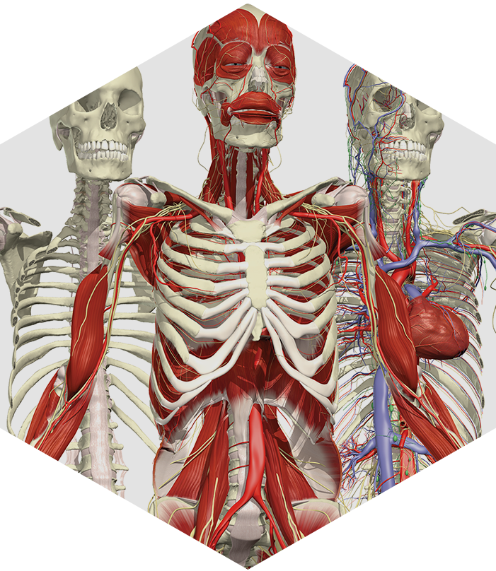
Primal Pictures develops its digital anatomical models using real scan and imaging data. Reviewed by qualified anatomists and external experts, these models give educators detailed resources for explaining structures, relationships, and movement.
A Foundation for Anatomy and Physiology Teaching
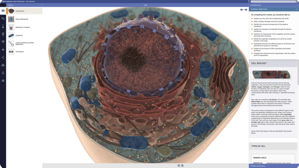
Make complex concepts easier to demonstrate with interactive 3D models, explanatory content, and learning activities. Give educators resources to reinforce instruction and help learners connect anatomical structures with their functions.
Make Patient Education More Visual
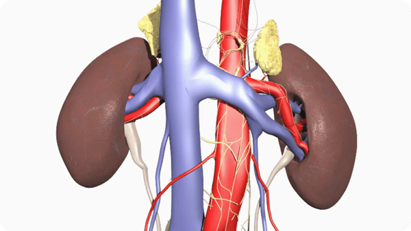
Use clear anatomical models to explain affected structures, conditions, and treatment plans. Give healthcare professionals visual resources to make conversations with patients easier to follow.
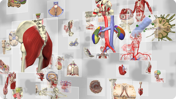
Anatomical Detail You Can Teach with Confidence
Primal Pictures combines real imaging data, anatomical expertise, and extensive review to create detailed digital models. Through Anatomy.tv, educators and learners can explore this content alongside clinical explanations, images, and related learning resources.
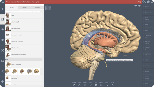
Bring Interactive Anatomy into Your Courses
Enrich lectures, course materials, and online learning with focused anatomical content. Use dynamic images and interactive resources to illustrate difficult concepts and encourage learners to explore beyond the classroom.
Interactive 3D Anatomy from Primal Pictures
Anatomy.tv brings together Primal Pictures’ anatomical models and resources for foundational sciences, clinical understanding, and professional learning. Educators can demonstrate complex relationships, while learners explore anatomy independently and revisit concepts throughout training and practice. TDS Health helps you identify the modules that fit your curricula, disciplines, and institutional learning needs.
Who Can Benefit From Primal Pictures?
-
EDUCATORS
Make Anatomy Easier to Teach and Apply
Explain structures, relationships, and movement with interactive 3D anatomy. Connect foundational concepts with clinical examples to help learners build understanding and prepare for practical application.
Enrich Your Teaching Materials
Use images and content in lectures, course materials, and online learning environments to reinforce your objectives and give learners additional ways to explore anatomy.
Extend Learning Beyond the Classroom
Provide web-based access for class preparation, independent study, and review. Help learners revisit concepts before laboratory sessions, assessments, and clinical training.

-
STUDENTS
Build a Strong Foundation in Anatomy and Physiology
Give students interactive resources to explore anatomy, prepare for laboratory sessions, and review for exams. Rotate models, add or remove layers, and select visible structures to learn more about their relationships and functions.
Flexible web-based access lets learners revisit content outside the classroom and continue building their understanding as coursework progresses.

-
Practitioners
Connect Anatomy with Clinical Practice
Give practitioners detailed anatomical references for professional review, teaching, and patient education. Explore structures and relationships relevant to clinical specialties, and use visual content to explain anatomical concepts during discussions of conditions and care.

-
HOSPITALS
Strengthen Hospital Education and Professional Learning
Bring interactive anatomy into staff education, clinical training, and patient communication. Choose resources that fit your departments and professional development priorities.
Refresh Anatomical Knowledge
Help healthcare teams revisit structures, functions, and clinical relationships through detailed models and explanatory content.
Make Clinical Explanations Clearer
Use visual resources to illustrate injuries, conditions, and treatment plans. Give professionals tools to explain affected anatomy and encourage informed conversations with patients.

-
-
Make Anatomy Easier to Teach and Apply
Explain structures, relationships, and movement with interactive 3D anatomy. Connect foundational concepts with clinical examples to help learners build understanding and prepare for practical application.
Enrich Your Teaching Materials
Use images and content in lectures, course materials, and online learning environments to reinforce your objectives and give learners additional ways to explore anatomy.
Extend Learning Beyond the Classroom
Provide web-based access for class preparation, independent study, and review. Help learners revisit concepts before laboratory sessions, assessments, and clinical training.
Build a Strong Foundation in Anatomy and Physiology
Give students interactive resources to explore anatomy, prepare for laboratory sessions, and review for exams. Rotate models, add or remove layers, and select visible structures to learn more about their relationships and functions.
Flexible web-based access lets learners revisit content outside the classroom and continue building their understanding as coursework progresses.
Connect Anatomy with Clinical Practice
Give practitioners detailed anatomical references for professional review, teaching, and patient education. Explore structures and relationships relevant to clinical specialties, and use visual content to explain anatomical concepts during discussions of conditions and care.
Strengthen Hospital Education and Professional Learning
Bring interactive anatomy into staff education, clinical training, and patient communication. Choose resources that fit your departments and professional development priorities.
Refresh Anatomical Knowledge
Help healthcare teams revisit structures, functions, and clinical relationships through detailed models and explanatory content.
Make Clinical Explanations Clearer
Use visual resources to illustrate injuries, conditions, and treatment plans. Give professionals tools to explain affected anatomy and encourage informed conversations with patients.
Explore Anatomy.tv
-
Overview
Primal Pictures Overview
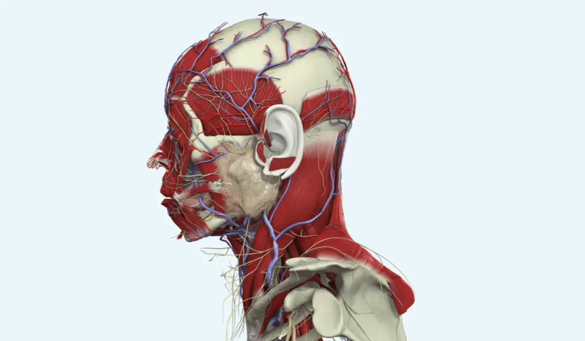
Primal Pictures’ anatomical models were developed using CT scans, MRI data, and cross-sectional cadaver images. Muscles, vessels, nerves, organs, and skeletal structures form a detailed digital representation of human anatomy.
Content is reviewed by qualified anatomists and external experts for each body area, giving educators a reliable foundation for teaching and clinical learning.
-
Key Features
Key Features for Teaching and Learning
- Explore detailed 3D anatomical structures.
- Rotate models 360 degrees and add or remove anatomical layers.
- Connect models with relevant text, dissections, clinical images, diagrams, videos, and imaging.
- Embed available images and content into websites, learning management systems, and virtual learning environments.
- Reinforce anatomy and physiology instruction across healthcare disciplines.
- Use anatomical references and images for professional review and patient education.
Primal Pictures Overview
Primal Pictures’ anatomical models were developed using CT scans, MRI data, and cross-sectional cadaver images. Muscles, vessels, nerves, organs, and skeletal structures form a detailed digital representation of human anatomy.
Content is reviewed by qualified anatomists and external experts for each body area, giving educators a reliable foundation for teaching and clinical learning.
Key Features for Teaching and Learning
- Explore detailed 3D anatomical structures.
- Rotate models 360 degrees and add or remove anatomical layers.
- Connect models with relevant text, dissections, clinical images, diagrams, videos, and imaging.
- Embed available images and content into websites, learning management systems, and virtual learning environments.
- Reinforce anatomy and physiology instruction across healthcare disciplines.
- Use anatomical references and images for professional review and patient education.
A Portfolio Built Around Your Anatomy Learning Needs
Primal Pictures’ models form the foundation of a broad portfolio available through Anatomy.tv. Choose resources that connect foundational study with imaging, clinical specialties, functional movement, and development.
-
Gross Anatomy
Gross Anatomy
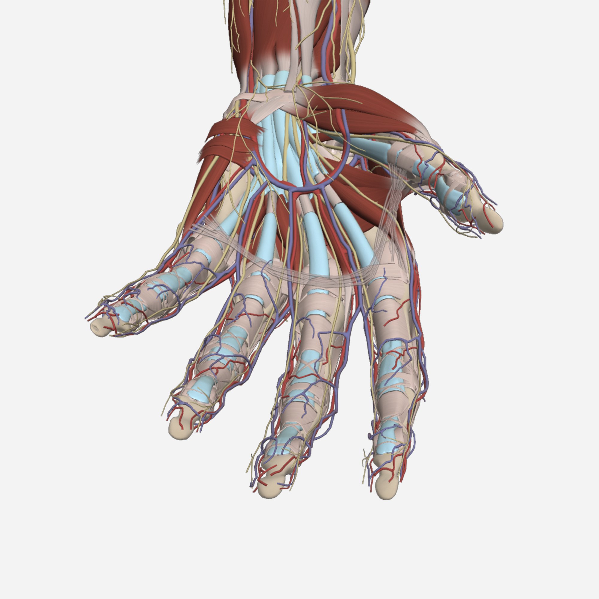
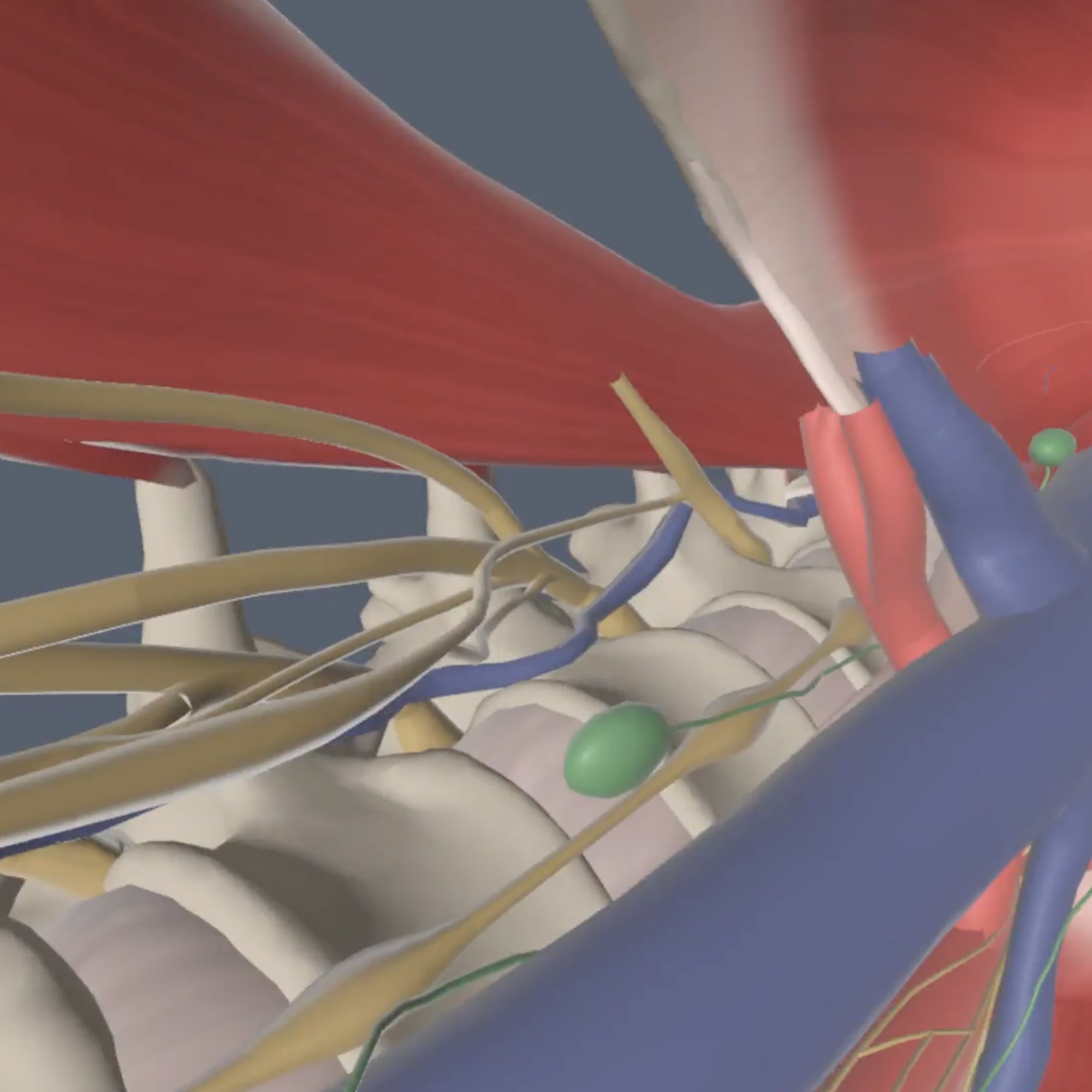
3D Real-time
Explore and virtually dissect 3D anatomy, create focused views, and test understanding with quizzes.
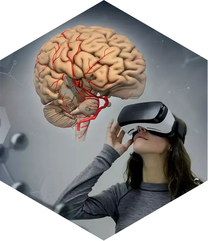
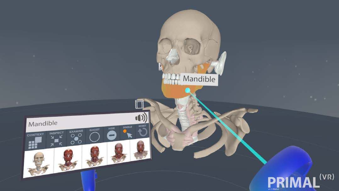
Primal VR
Give learners opportunities to explore and interact with anatomical models in virtual reality.
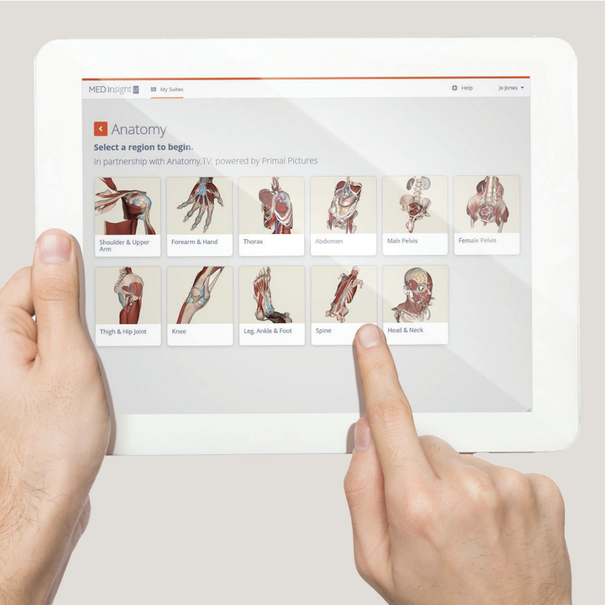
3D Atlas
Guide learning through regional 3D models, an image bank, and detailed explanatory text.
PALMs
Perceptive and Adaptive Learning Modules assess anatomical knowledge and adapt practice to each learner.
-
Clinical & Imaging
Clinical & Imaging
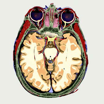
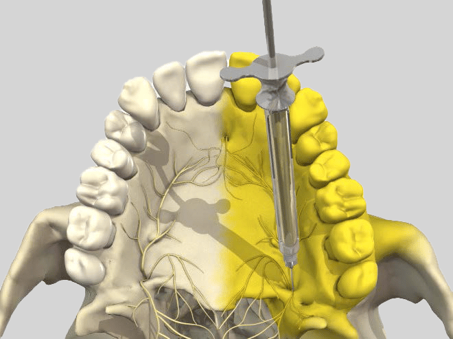
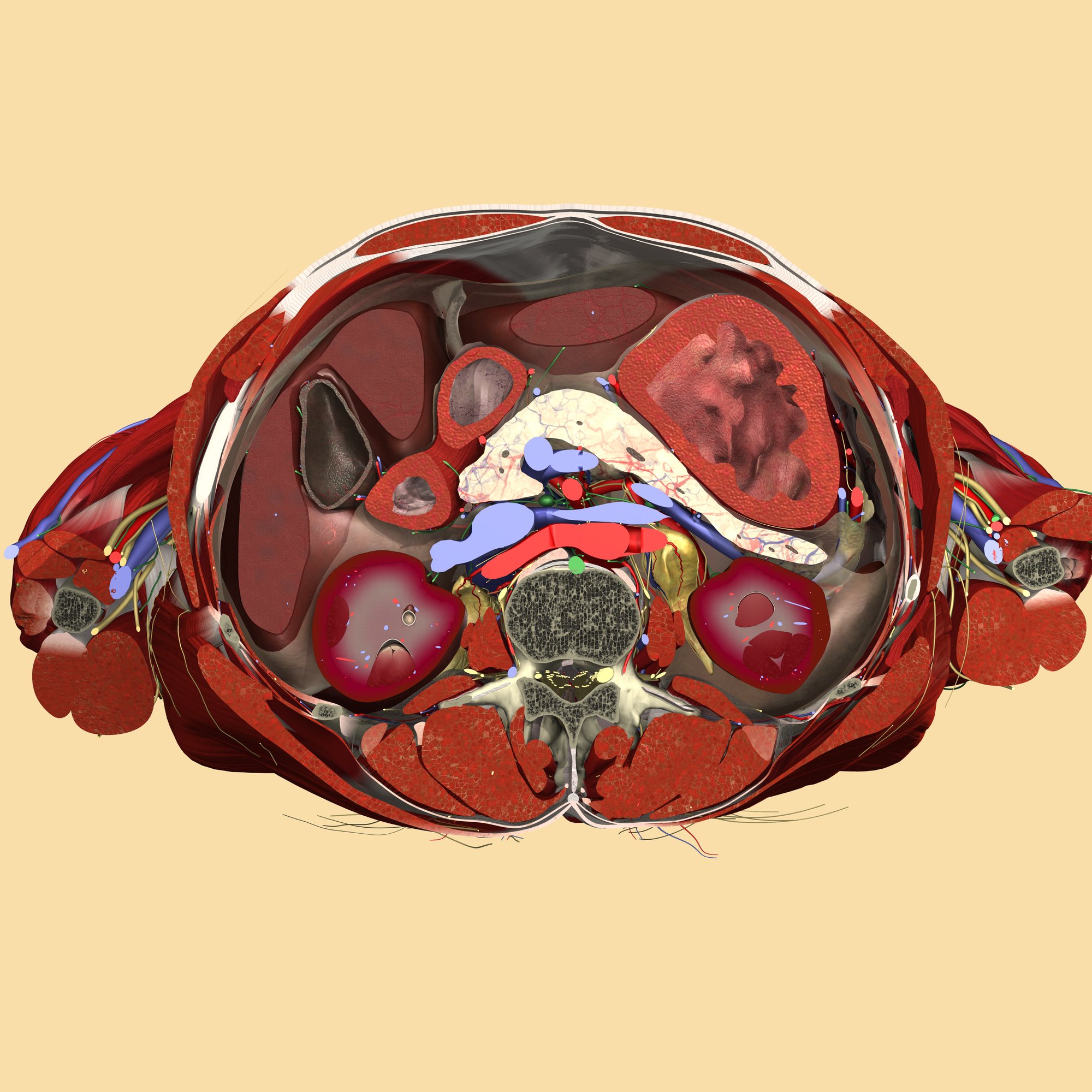
Disease & Conditions
Illustrate how disease affects anatomy with expert-reviewed 3D models and videos.
Clinical Specialties
Connect anatomy with clinical learning in dentistry, pelvic floor disorders, speech-language pathology, and audiology.
Imaging
Explore radiology and cross-sectional anatomy through resources covering ultrasound of the limbs and cross-sectional anatomy of the trunk.
-
Anatomy & Physiology
Anatomy & Physiology
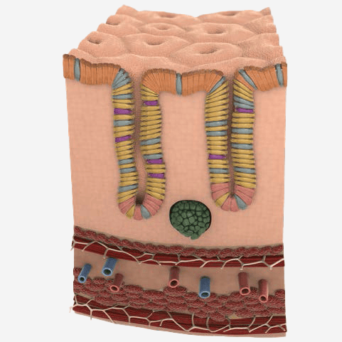
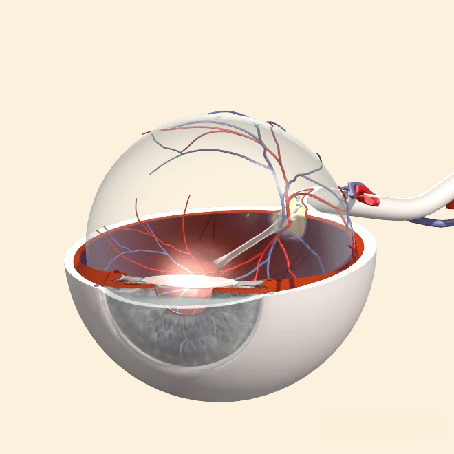
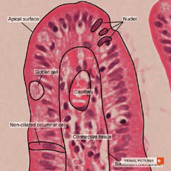
Anatomy & Physiology
Teach foundational concepts through 20 systems-based modules with guided text, 3D models, videos, quizzes, and case studies.
-
Functional Anatomy
Functional Anatomy
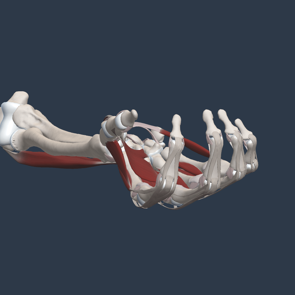
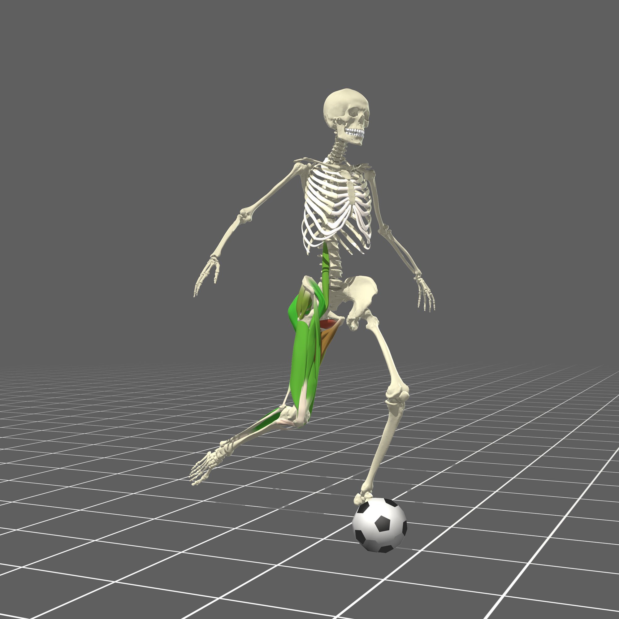
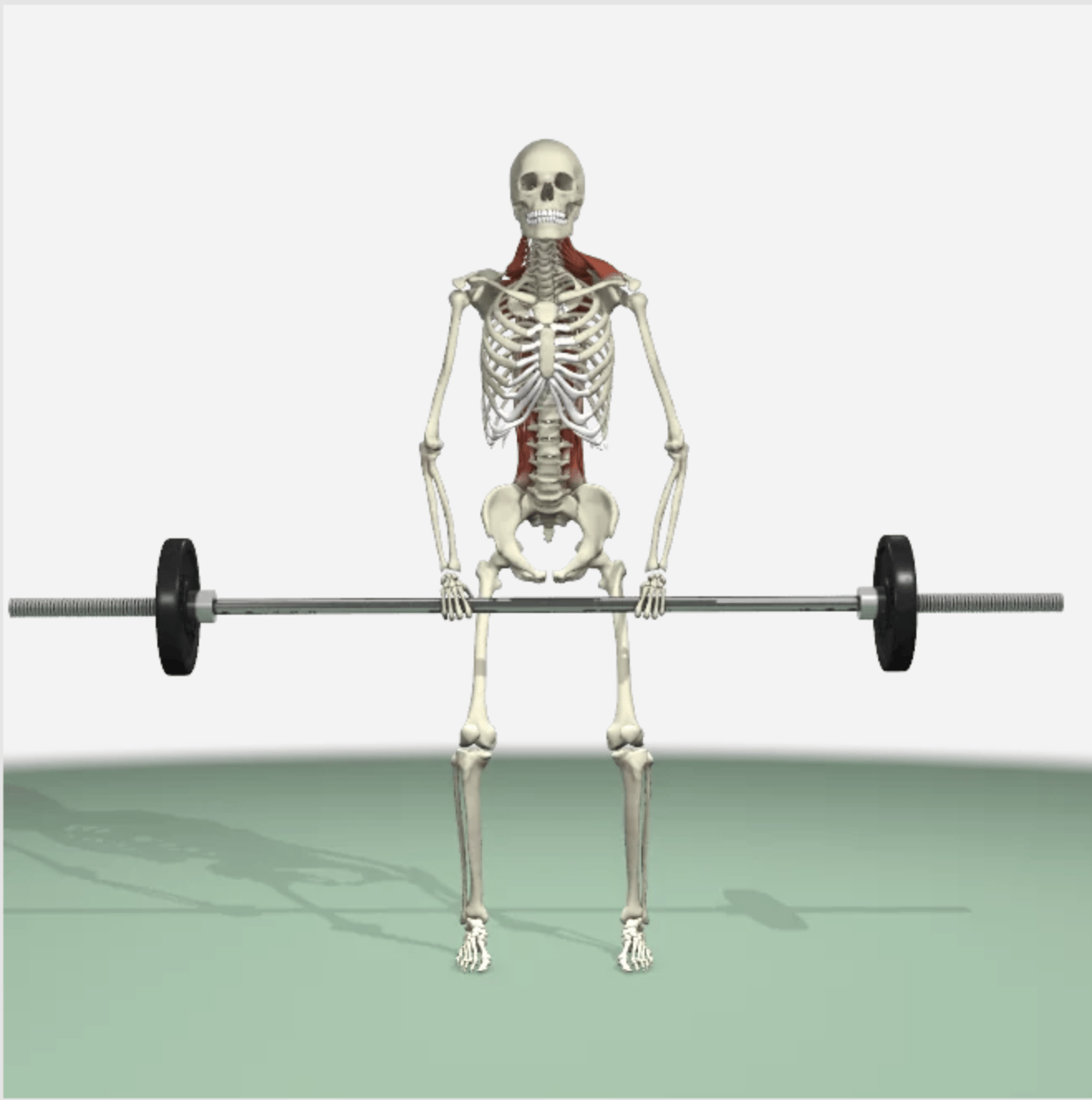
Real-Time Functional Anatomy
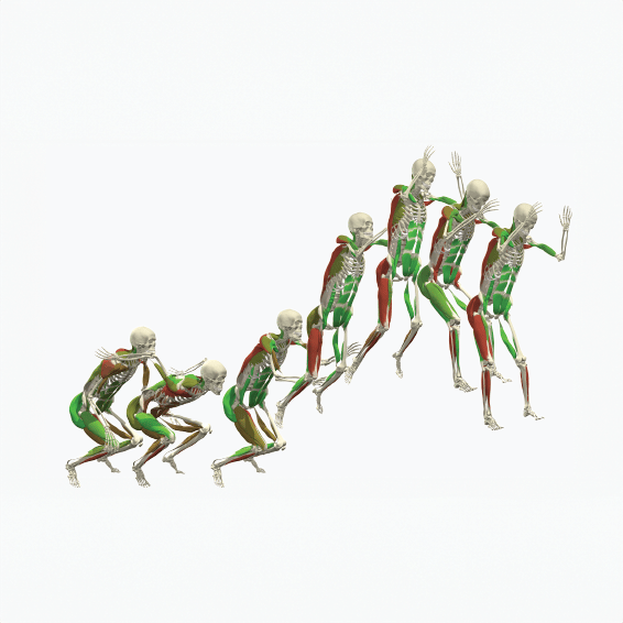
Demonstrate functional movements and goniometry techniques with an interactive 3D model in motion.
Functional Anatomy
Connect anatomical structures with movement through guided animations and 3D models.
Resistance Training
Explore relevant anatomy and demonstrate muscle function through exercise animations.
-
Embryology
Embryology
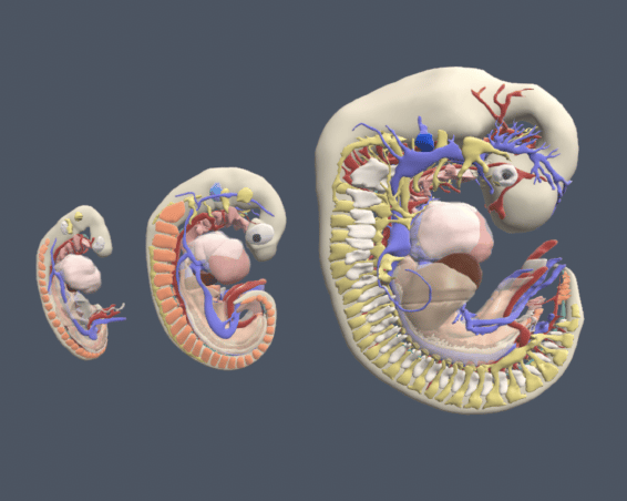
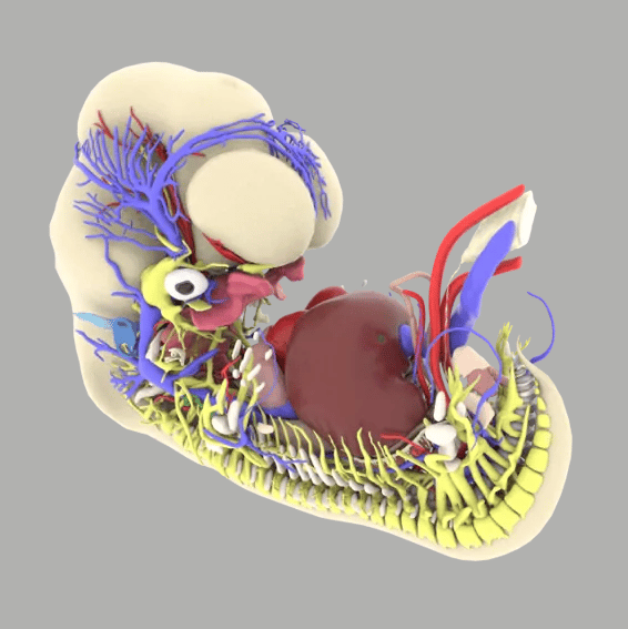
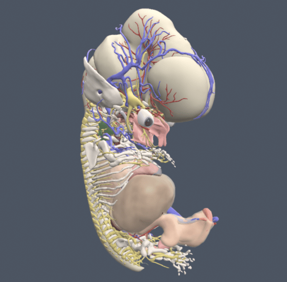
Real-time Embryology
Explore development in 3D, with tools to rotate models, examine structures, and create focused views.
-
Anatomy Learning Outcomes for Medicine
Anatomy Learning Outcomes for Medicine
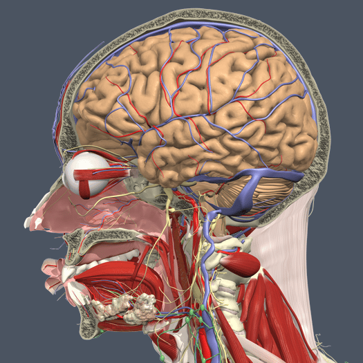
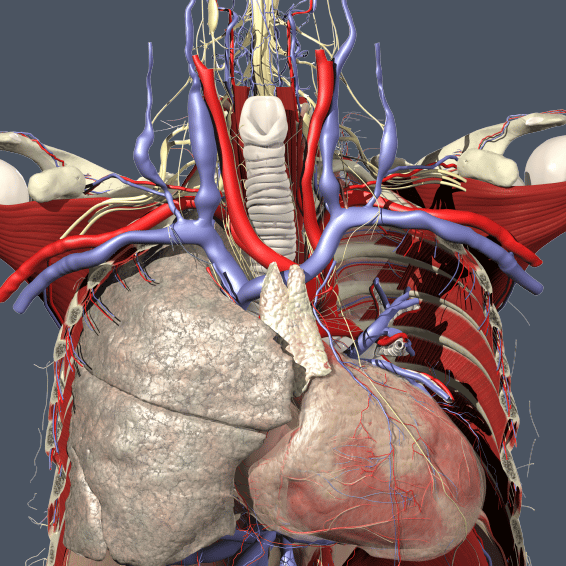
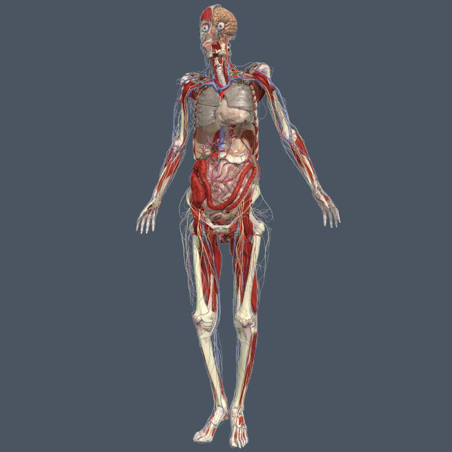
Anatomy Learning Outcomes for Medicine
Access videos from more than 50 anatomy experts explaining the 156 Anatomical Society learning outcomes for medicine. Available with subscriptions to Gross Anatomy, Clinical & Imaging, and Anatomy & Physiology.
-
Anatomy Tutorials for Physical Therapy
Anatomy Tutorials for Physical Therapy
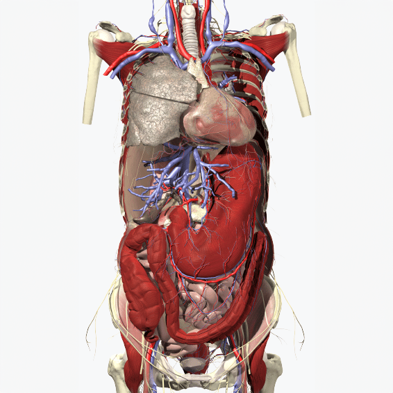
Anatomy Tutorials for Physical Therapy
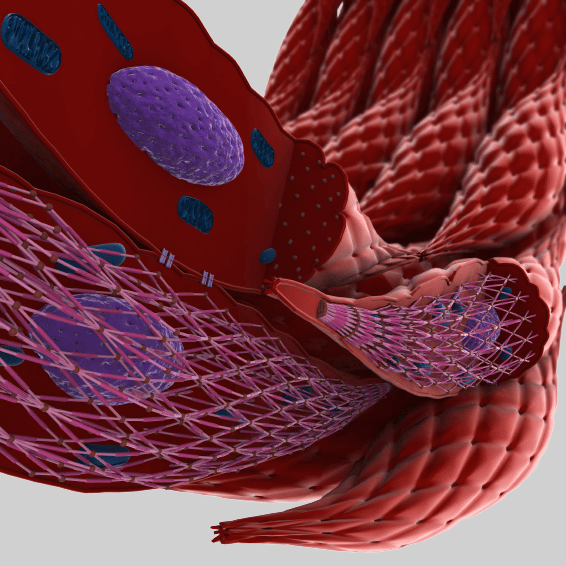
Explore videos from Physiopedia experts explaining key concepts in musculoskeletal anatomy. Available with subscriptions to Clinical & Imaging, Anatomy & Physiology, and Functional Anatomy.
Gross Anatomy
3D Real-time
Explore and virtually dissect 3D anatomy, create focused views, and test understanding with quizzes.
Primal VR
Give learners opportunities to explore and interact with anatomical models in virtual reality.
3D Atlas
Guide learning through regional 3D models, an image bank, and detailed explanatory text.
PALMs
Perceptive and Adaptive Learning Modules assess anatomical knowledge and adapt practice to each learner.
Clinical & Imaging
Disease & Conditions
Illustrate how disease affects anatomy with expert-reviewed 3D models and videos.
Clinical Specialties
Connect anatomy with clinical learning in dentistry, pelvic floor disorders, speech-language pathology, and audiology.
Imaging
Explore radiology and cross-sectional anatomy through resources covering ultrasound of the limbs and cross-sectional anatomy of the trunk.
Anatomy & Physiology
Anatomy & Physiology
Teach foundational concepts through 20 systems-based modules with guided text, 3D models, videos, quizzes, and case studies.
Functional Anatomy
Real-Time Functional Anatomy
Demonstrate functional movements and goniometry techniques with an interactive 3D model in motion.
Functional Anatomy
Connect anatomical structures with movement through guided animations and 3D models.
Resistance Training
Explore relevant anatomy and demonstrate muscle function through exercise animations.
Embryology
Real-time Embryology
Explore development in 3D, with tools to rotate models, examine structures, and create focused views.
Anatomy Learning Outcomes for Medicine
Anatomy Learning Outcomes for Medicine
Access videos from more than 50 anatomy experts explaining the 156 Anatomical Society learning outcomes for medicine. Available with subscriptions to Gross Anatomy, Clinical & Imaging, and Anatomy & Physiology.
Anatomy Tutorials for Physical Therapy
Anatomy Tutorials for Physical Therapy
Explore videos from Physiopedia experts explaining key concepts in musculoskeletal anatomy. Available with subscriptions to Clinical & Imaging, Anatomy & Physiology, and Functional Anatomy.

There is a digital healthcare revolution underway that is fundamentally changing the way medical information is shared, presented and used by physicians, educators and students. At the heart of this revolution is Anatomy.tv, the World’s First Complete 3D Model of Human Anatomy.

We are constantly looking for additional information which would help our students. Not everybody learns the same way, so there would be additional ancillary help outside the classroom the student could access himself or herself that would be sort of a programmed learning experience for them.

The 3D real-time viewer I was pleased to use is AMAZING!! It's so easy to use and so beautiful. My colleagues were completely surprised by the astonishing quality and simplicity in which it is to use. As a radiologist we use images all day, every day. Your images are THE images to use in presentations or just for anatomic correlation during evaluation of scans.

Anatomy.tv offers a wide array of anatomical images, a growing library of pathological anatomical images, and excellent videos on many topic areas. I utilize Anatomy.tv regularly for our continuing education courses on PhysicalTherapy.com. I work with our presenters and other colleagues at continued.com to identify the appropriate images for their presentations which in turn assists our learners to understand and apply the content taught by our expert presenters in their respective fields.

I took a look, I saw Primal’s Embed feature, and I was like, sold! I like how quick and easy it is to enrich my course materials using the same trusted content I've always used on Anatomy.tv. I just saw that it said, Embed, and then you can go choose Canvas. As the embedded content retains its original dynamic functionality, my students can rotate, layer, highlight, and use the labeling for interactive images or simply play, pause, and replay the videos. One of the big things is that they just really enjoy the amount of material that they have to study with. And for everything that we’re able to give to these students, it has definitely improved their performance.